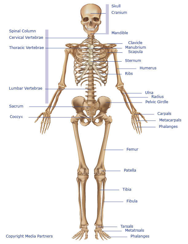
Lower Body Bone Diagram / Divisions Of The Skeletal System Anatomy And Physiology. Free homeostasis and the human body worksheets 2021 The femur (thigh bone), tibia and fibula (lower leg bones), clavicle (collar bone), humerus (upper arm bone), the radius and the ulna (lower arm), metacarpals (hand bones), metatarsals (foot bones) and phalanges (finger and toe bones). The bones together make up the hip. The bones of the leg are the femur, tibia, fibula and patella.the foot bones shown in this diagram are the talus, navicular, cuneiform, cuboid, metatarsals and calcaneus. In the back and elsewhere in the body, tendons attach muscles to bones.
These human anatomy worksheets serve as a nice intro to physiology for kids. The patella and the pisiform bone of the carpals are the only sesamoid bones that are counted as part of the 206 bones of the body. The bones of the legs are those that make up the thigh, the lower half of the legs, and the feet. Diagrams of human muscles lower arm muscles diagram human muscle. Bone long blood diaphysis vector anatomical anatomy articular biology body calcium cartilage cell compact detail diagram education educational endosteum epiphysis forelimb.
Horse Anatomy Mobility Health from cdn.shopify.com Your lower back (lumbar spine) is the anatomic region between your lowest rib and the upper part of the buttock. The bones of the leg are the femur, tibia, fibula and patella.the foot bones shown in this diagram are the talus, navicular, cuneiform, cuboid, metatarsals and calcaneus. The vertebral column of the lower back includes the five lumbar vertebrae, the sacrum, and the coccyx. Free trial, examples, and templates. This diagram depicts picture of female reproductive system diagram 1024×1204 with parts and labels. How about the muscles of the head. Human bone anatomy diagram 8 photos of the human bone anatomy diagram female pelvic bone anatomy diagram, hip bone anatomy diagram, human body anatomy diagram, human bone anatomy 3d, human bone anatomy game, human bone anatomy quiz, human muscle anatomy, leg bone anatomy diagram, human anatomy, female pelvic bone anatomy diagram, hip bone. Female anatomy includes the external genitals, or the vulva, and the internal reproductive organs.
Test your knowledge of the muscles of the face.
Human bone anatomy diagram 8 photos of the human bone anatomy diagram female pelvic bone anatomy diagram, hip bone anatomy diagram, human body anatomy diagram, human bone anatomy 3d, human bone anatomy game, human bone anatomy quiz, human muscle anatomy, leg bone anatomy diagram, human anatomy, female pelvic bone anatomy diagram, hip bone. Key bones in the abdominal area include the base of the ribcage and the lumbar spine in the lower back. The bones of the lower limbs. Can you name the muscles of the body from the side?. A muscle's origin is where a tendon attaches it to the *less* movable bone. ☛ when you wave your wrist, all the bones of your arm are at work. This diagram depicts picture of female reproductive system diagram 1024×1204 with parts and labels. Learn the muscles of the arm. The patella and the pisiform bone of the carpals are the only sesamoid bones that are counted as part of the 206 bones of the body. Free trial, examples, and templates. The human skeleton is the internal framework of the human body. The bones together make up the hip. Learn how to communicate up to six times better by using visuals with smartdraw.
Free trial, examples, and templates. In all, there are believed to be 80 organs in your body, all serving different functions and uses. Female lower body parts diagram ~ diagram from i.pinimg.com i made this basic guide for male and female body proportions according to andrew loomis for myself, but download diagram showing anatomy of human body vector art. This article looks at the anatomy of the back, including bones, muscles. At the level of the pelvic bones, the abdomen.
Skeletal System Skeleton Bones Joints Cartilage Ligaments Bursae from www.healthpages.org The bmi is defined as the body mass divided by the square of the body height. The bones of the lower limbs. Free homeostasis and the human body worksheets 2021 Balance the weight of your head on top of your spine. The human skeleton is the internal framework of the human body. These human anatomy worksheets serve as a nice intro to physiology for kids. The hip itself is a ball and socket joint, much like the shoulder.the structures necessary to create this joint are the socket, the joint capsule, muscle, ligaments, and the neck. The back supports the weight of the body, allowing for flexible movement while protecting vital organs and nerve structures.
Free trial, examples, and templates.
On the other hand, the insertion is where a tendon attaches that muscle to the *more* movable bone. This diagram depicts picture of female reproductive system diagram 1024×1204 with parts and labels. The bones together make up the hip. 1 your spine in this region has a natural inward curve. Can you name the muscles of the body from the side?. This is the longest bone in the human body, and is also known as the thigh bone. The bones of the hip include the femur, the ilium, the ischium, and the pubis. The male reproductive system includes the following structures. Human bone anatomy diagram 8 photos of the human bone anatomy diagram female pelvic bone anatomy diagram, hip bone anatomy diagram, human body anatomy diagram, human bone anatomy 3d, human bone anatomy game, human bone anatomy quiz, human muscle anatomy, leg bone anatomy diagram, human anatomy, female pelvic bone anatomy diagram, hip bone. Diagrams of human muscles lower arm muscles diagram human muscle. The femur is the only bone of the thigh. The vertebral column of the lower back includes the five lumbar vertebrae, the sacrum, and the coccyx. This article looks at the anatomy of the back, including bones, muscles.
Posted on june 7, 2016 by admin. Diagrams of human muscles lower arm muscles diagram human muscle. The hip itself is a ball and socket joint, much like the shoulder.the structures necessary to create this joint are the socket, the joint capsule, muscle, ligaments, and the neck. This article looks at the anatomy of the back, including bones, muscles. How about the muscles of the head.
Stock Image An Anterior View Of The Bones Of The Lower Body The Surfaceanatomy Of The Body Is Semi Transparent And Tinted Red 109772 01axwrnw 3d4medical Search Medical Scientific Stock from www.medicalimages.com In the back and elsewhere in the body, tendons attach muscles to bones. The patella and the pisiform bone of the carpals are the only sesamoid bones that are counted as part of the 206 bones of the body. The human skeleton is the internal framework of the human body. Get product information, training, support, solutions and more. Human body homepage the body homepage interactive body skeleton game facts and features skeleton anatomy diagram arm and shoulder broken bones hands and feet joints pelvis ribcage skull spine teeth psychology tests disgust spot the fake smiles memory test. The bones of the lower limbs. Did you know… ☛ the femur bone, also known as the thigh bone, is the longest bone in the body. The back supports the weight of the body, allowing for flexible movement while protecting vital organs and nerve structures.
It is deemed far stronger than concrete!
The hip itself is a ball and socket joint, much like the shoulder.the structures necessary to create this joint are the socket, the joint capsule, muscle, ligaments, and the neck. Diagrams of human muscles lower arm muscles diagram human muscle. The diaphragm forms the upper surface of the abdomen. The human skeleton is the internal framework of the human body. Downloadable pdf anatomy worksheets can be printed, labeled and colored to practice your understanding of human anatomy and physiology. This article looks at female body parts and their functions, and it provides an interactive diagram. The vertebral column of the lower back includes the five lumbar vertebrae, the sacrum, and the coccyx. The femur (thigh bone), tibia and fibula (lower leg bones), clavicle (collar bone), humerus (upper arm bone), the radius and the ulna (lower arm), metacarpals (hand bones), metatarsals (foot bones) and phalanges (finger and toe bones). Bone diagram forehead (frontal bone) nose bones (nasals) cheek bone (zygoma) upper jaw (maxilla) lower jaw (mandible) breast bone (sternum) upper arm bone (humerus) lower arm bone (ulna) thigh bone (femur) collar bone (clavicle) toe bones (phalanges) ankle bones. These are the body's levers, they allow movement, particularly in the limbs e.g. The bones of the hip include the femur, the ilium, the ischium, and the pubis. Daniel nelson on june 5, 2018 8 comments ! Because of the important organs situated in the abdominal area, many health concerns stem.
The long bones of the body contain many distinct regions due to the way in which they develop lower body diagram. Because of the important organs situated in the abdominal area, many health concerns stem.
Share :
Post a Comment
for "Lower Body Bone Diagram / Divisions Of The Skeletal System Anatomy And Physiology"


Post a Comment for "Lower Body Bone Diagram / Divisions Of The Skeletal System Anatomy And Physiology"